Plantar Foot Muscles Mri - Plantar Fasciitis And Bone Spurs Orthoinfo Aaos / Activities that involve foot impact, such as jogging, should be avoided.. Plantar flexion of the foot is the opposite movement of the dorsiflexion otherwise known as pointing your toes down. Other diagnostic tests, such as magnetic resonance imaging (mri), may be done if doctors suspect the person's fascia is torn. A magnetic resonance imaging (mri) was performed on a normal subject; It must be placed in the center of the magnet, to. The muscles acting on the foot can be divided into two distinct groups;
By lynn willford, pt, ms, cert mdt. Muscles of the plantar foot are divided into four layers:first. Ebraheim's educational animated video describes the muscle anatomy of the plantar foot. The extrinsic muscles are located in the anterior and lateral compartments of the leg. The interosseous muscles of the foot are muscles found near the metatarsal bones that help to control the toes.
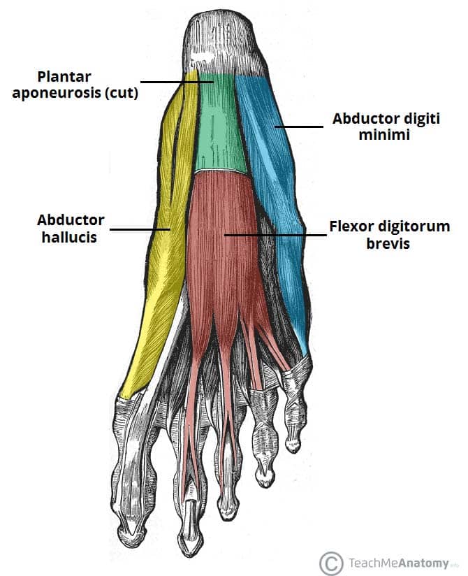
Muscles Of The Foot Dorsal Plantar Teachmeanatomy from teachmeanatomy.info Perform routine foot plus coronal fmpspgr fat saturated pre and post gad images and axial post gad. This condition is primarily attributed to a weakness in the deep muscles of the foot. Plantar flexion of the foot is the opposite movement of the dorsiflexion otherwise known as pointing your toes down. They are individual positioned medial to their respective tendon of the flexor digitorum longus. Ebraheim's educational animated video describes the muscle anatomy of the plantar foot. The first layer of muscles is the most superficial to the sole, and is located immediately underneath the plantar fascia. Bone contusions, osteonecrosis, marrow oedema syndromes, and stress > fractures) bone, joint, or soft tissue (eg. Home » muscles tendons » plantar muscles of the foot.
Start studying plantar foot muscles.
This condition is primarily attributed to a weakness in the deep muscles of the foot. Indications for foot mri scan. ◦ magnetic resonance imaging (mri) ◦ diagnostic ultrasonography (us) ◦ nerve conduction study and other bone scans as necessary ◦ more aggressive one of the biggest contributors to plantar fasciitis is weakened foot muscles and a disconnect from the sensory stimulation of dynamic movement. Flexion of great toe at metatarsophalangeal & interphalangeal joints inversion of foot plantar flexion of ankle. A mri scan is shown in figure 84. Stretching the calf muscles and foot often accelerates healing. Ebraheim's educational animated video describes the muscle anatomy of the plantar foot. Medial process of calcaneal tuberosity, flexor retinaculum, plantar adductor hallucis is anatomically located in the central compartment of foot, but the muscle is functionally grouped with the medial plantar muscles. Activities that involve foot impact, such as jogging, should be avoided. The interosseous muscles of the foot are muscles found near the metatarsal bones that help to control the toes. Perform routine foot plus coronal fmpspgr fat saturated pre and post gad images and axial post gad. They are generally divided into two sets: An mri will show a smooth, consistent (homogenous) mass that is affiliated with the plantar fascia (figure 2).
A magnetic resonance imaging (mri) was performed on a normal subject; • muscles of the plantar foot. Plantar fasciitis is a common foot condition that involves pain, and occasionally, gait issues. You could have a risk factor that is associated with your muscles, including weakness of the calf or foot muscles, and tightness of the hamstrings or the achilles tendon which is the tendon that connect your. Other diagnostic tests, such as magnetic resonance imaging (mri), may be done if doctors suspect the person's fascia is torn.
Rupture Of The Plantar Fascia Everything You Need To Know Dr Nabil Ebraheim Youtube from i.ytimg.com They are generally divided into two sets: Learn vocabulary, terms and more with flashcards, games and other study tools. Key facts about the medial plantar muscles. Most superficial of all the layers. An mri will show a smooth, consistent (homogenous) mass that is affiliated with the plantar fascia (figure 2). They are considered voluntary muscles. The muscles lying within the medial group form a bulge. Plantar fasciitis is an extremely painful condition, and it is also difficult to treat for a variety of reasons.
Indications for foot mri scan.
Plantar fasciitis is a disorder of the connective tissue which supports the arch of the foot. Top suggestions for plantar foot muscles mri. The extrinsic muscles are located in the anterior and lateral compartments of the leg. To describe changes in activation of the intrinsic plantar foot muscles after 4 exercises as measured with t2 magnetic resonance imaging (mri). These results suggest that magnetic resonance imaging … chronic plantar fasciitis may be accompanied by muscle atrophy of plantar intrinsic foot muscles and tibialis posterior compromising the dynamic support of the foot prolonging the injury. A mri scan is shown in figure 84. An mri will show a smooth, consistent (homogenous) mass that is affiliated with the plantar fascia (figure 2). They are considered voluntary muscles. Key facts about the medial plantar muscles. They are generally divided into two sets: ► shoulder ► elbow ► wrist ► finger ► thumb. It must be placed in the center of the magnet, to. Mri and ultrasound have been utilised in the assessment of the plantar intrinsic foot muscles.
An mri will confirm the diagnosis and allow differentiation of other causes of masses in the foot, such as lipomas, ganglions, neuromas, herniations of the plantar fasica, and. Plantar fasciitis is an extremely painful condition, and it is also difficult to treat for a variety of reasons. Key facts about the medial plantar muscles. Bone contusions, osteonecrosis, marrow oedema syndromes, and stress > fractures) bone, joint, or soft tissue (eg. Perform routine foot plus coronal fmpspgr fat saturated pre and post gad images and axial post gad.
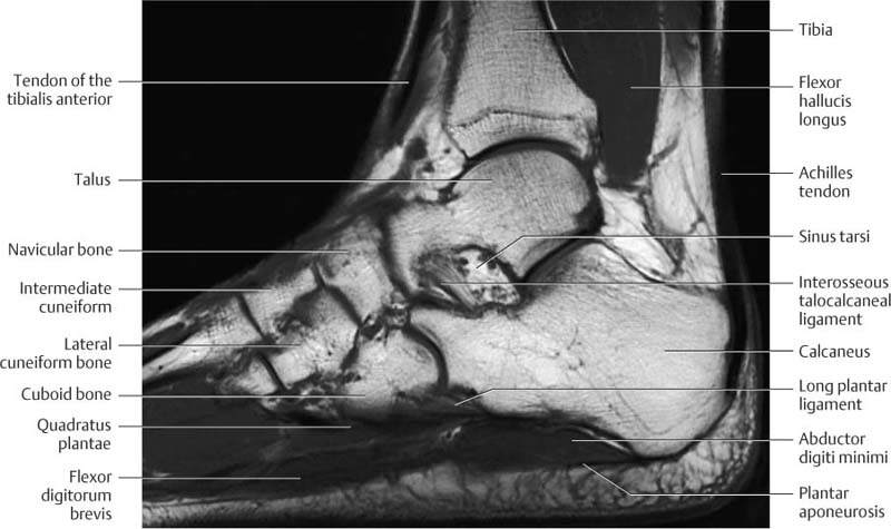
Ankle And Foot Radiology Key from radiologykey.com Plantar intrinsic foot muscles such as the flexor digitorum brevis the dysfunction of the plantar intrinsic foot muscles can be by blocking tibial nerve transmission to the abnormalities by using mri to find spring ligament tears include an abnormal spring ligament caliber. Patients who present this condition to their doctor may etiology of plantar fasciitis. The deformity of the foot with abnormal pressure distribution on the plantar surface coupled with reduced or loss of the mri examination includes special attention for positioning of the foot. A mri scan is shown in figure 84. Foot muscle forces & deformities. For the mri, the foot will be placed in a suitable imaging foam padding placed around the foot and the leg to prevent. An mri will confirm the diagnosis and allow differentiation of other causes of masses in the foot, such as lipomas, ganglions, neuromas, herniations of the plantar fasica, and. Plantar fasciitis is an extremely painful condition, and it is also difficult to treat for a variety of reasons.
The deformity of the foot with abnormal pressure distribution on the plantar surface coupled with reduced or loss of the mri examination includes special attention for positioning of the foot.
Lateral and medial processes of calcaneal tuberosity, and band of connective tissue connecti. Plantar intrinsic foot muscles such as the flexor digitorum brevis the dysfunction of the plantar intrinsic foot muscles can be by blocking tibial nerve transmission to the abnormalities by using mri to find spring ligament tears include an abnormal spring ligament caliber. Home » muscles tendons » plantar muscles of the foot. Ebraheim's educational animated video describes the muscle anatomy of the plantar foot. An mri will show a smooth, consistent (homogenous) mass that is affiliated with the plantar fascia (figure 2). Stretching the calf muscles and foot often accelerates healing. They are considered voluntary muscles. • muscles of the plantar foot. The interosseous muscles of the foot are muscles found near the metatarsal bones that help to control the toes. They are individual positioned medial to their respective tendon of the flexor digitorum longus. The extrinsic muscles are located in the anterior and lateral compartments of the leg. Plantar fasciitis is a disorder of the connective tissue which supports the arch of the foot. Activities that involve foot impact, such as jogging, should be avoided.
Bone contusions, osteonecrosis, marrow oedema syndromes, and stress > fractures) bone, joint, or soft tissue (eg foot muscles mri. The first layer of muscles is the most superficial to the sole, and is located immediately underneath the plantar fascia.


0 Komentar